File list
This special page shows all uploaded files.
| Date | Name | Thumbnail | Size | User | Description | Versions |
|---|---|---|---|---|---|---|
| 04:13, 21 August 2013 | IPLab10Crypto12.jpg (file) |  |
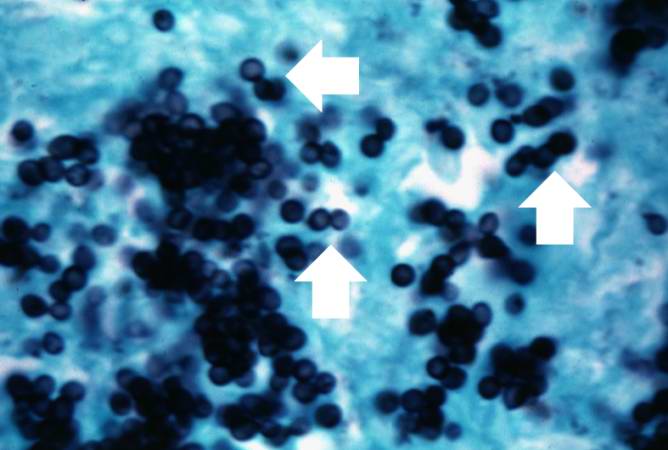
43 KB | Seung Park | This is a touch prep of fresh lung tissue that was allowed to air dry and then stained to show the mucopolysaccharide capsule around the cryptococcal organisms (arrows). | 1 |
| 04:12, 21 August 2013 | IPLab10Crypto11.jpg (file) |  |
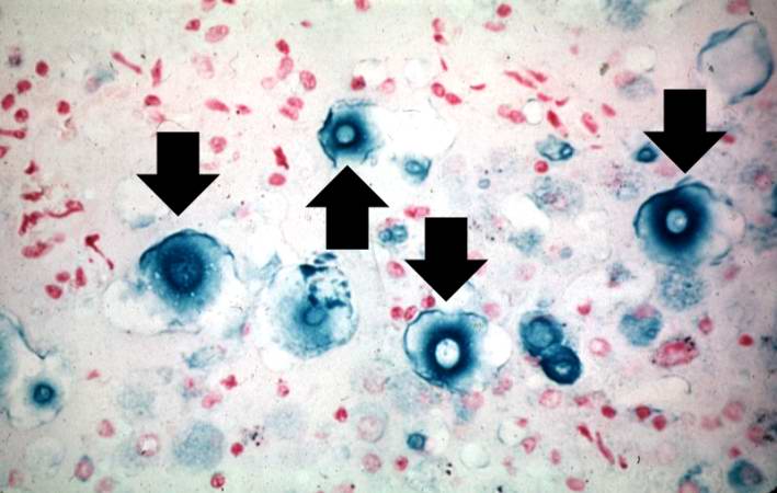
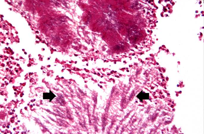
61 KB | Seung Park | This is a higher-power photomicrograph of lung section stained with Alcian blue. The mucopolysaccharide capsule shrinks during processing with this stain, thereby producing a shrunken central appearance with the formation of spikes around each organism. | 1 |
| 04:12, 21 August 2013 | IPLab10Crypto10.jpg (file) |  |
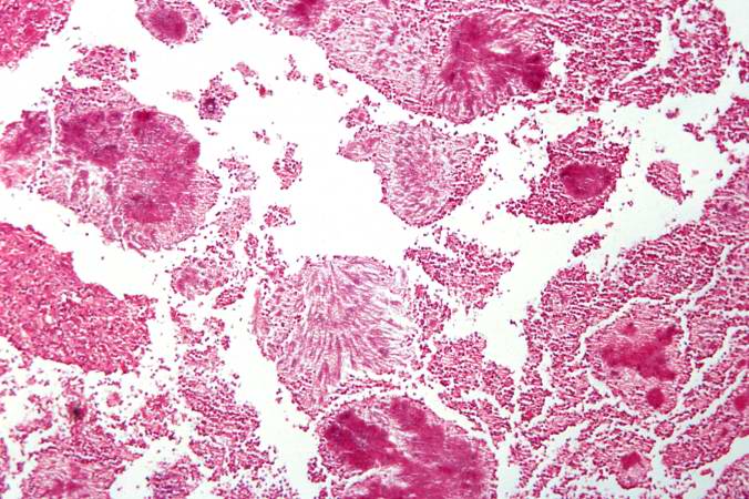
84 KB | Seung Park | This is a low-power photomicrograph of lung section stained with Alcian blue, which stains the acidic glycosaminoglycans making up the coat of the cryptococcal organism. | 1 |
| 04:12, 21 August 2013 | IPLab10Crypto9.jpg (file) |  |
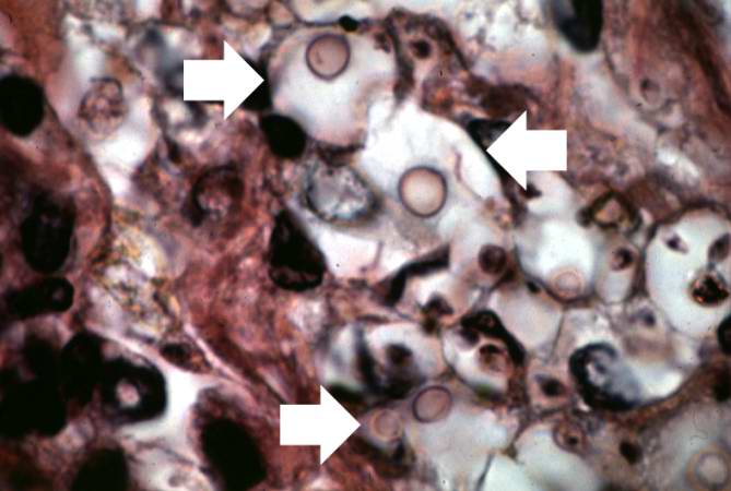
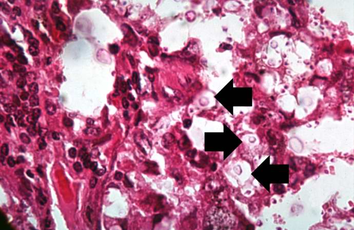
39 KB | Seung Park | This higher-power photomicrograph of a cryptococcal organism shows more clearly the nucleus surrounded by the large extracellular capsule (arrows). | 1 |
| 04:12, 21 August 2013 | IPLab10Crypto8.jpg (file) |  |
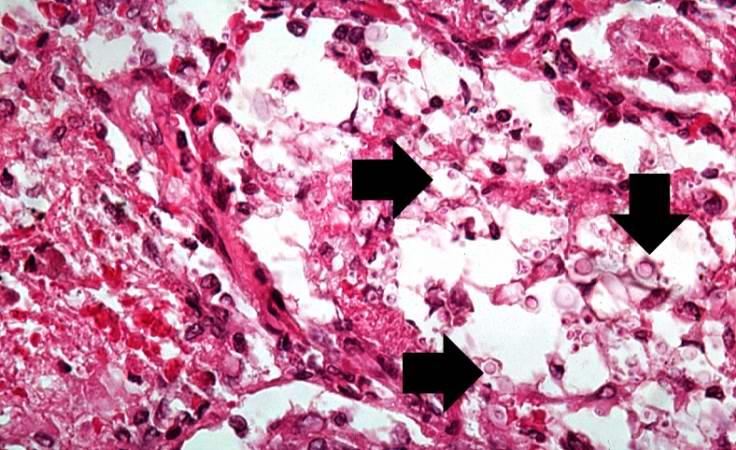
55 KB | Seung Park | Cryptococcal organisms can also be seen in this high-power photomicrograph of the cryptococcal lesion. Some of the organisms have a well-defined halo (arrows) due to the mucopolysaccharide coat which surrounds them. | 1 |
| 04:11, 21 August 2013 | IPLab10Crypto7.jpg (file) |  |
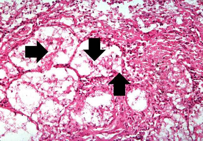
75 KB | Seung Park | This is another high-power photomicrograph of the cryptococcal lesion. In this section, numerous cryptococcal organisms (5-10 mm in diameter) can be seen (arrows). Note that there is very little inflammatory reaction. | 1 |
| 04:11, 21 August 2013 | IPLab10Crypto6.jpg (file) |  |
77 KB | Seung Park | This is a high-power photomicrograph of the cryptococcal lesion. Some of the organisms have been expelled during processing, but some cryptococcal organisms can be seen (arrows). | 1 |
| 04:11, 21 August 2013 | IPLab10Crypto5.jpg (file) |  |
71 KB | Seung Park | This is a higher-power photomicrograph of the cryptococcal lesion. The air spaces are filled with organisms (arrows). | 1 |
| 04:11, 21 August 2013 | IPLab10Crypto4.jpg (file) |  |
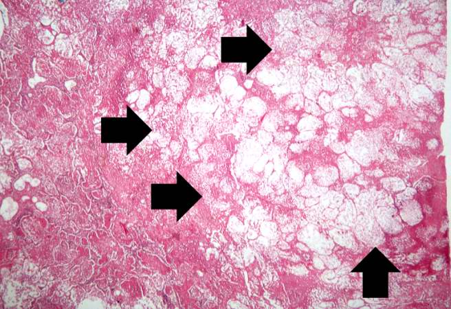
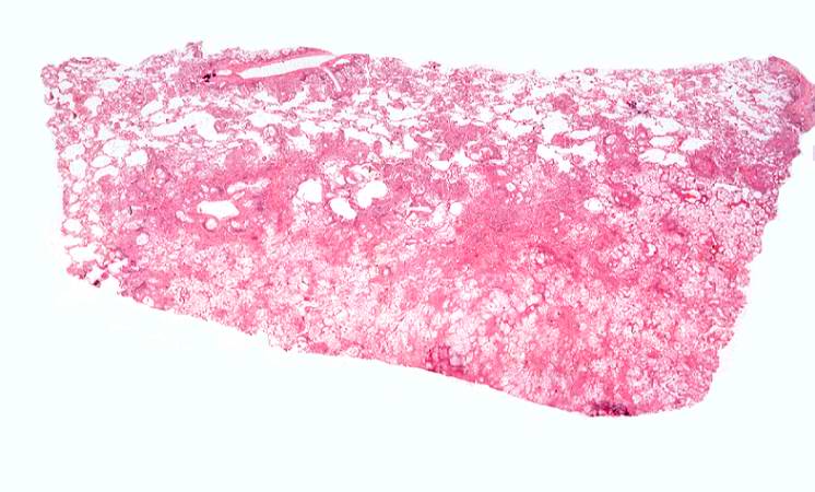
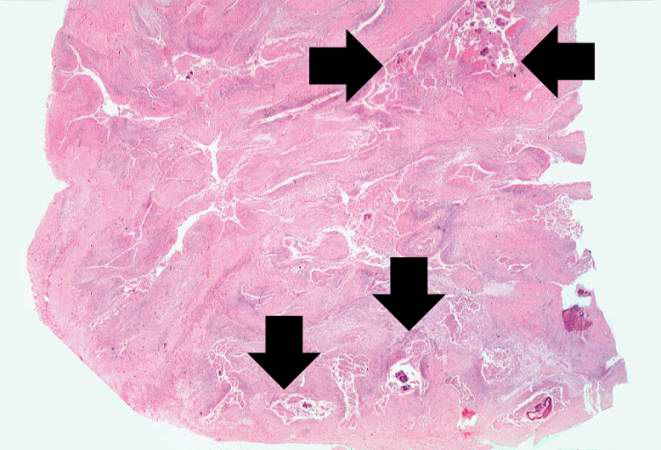
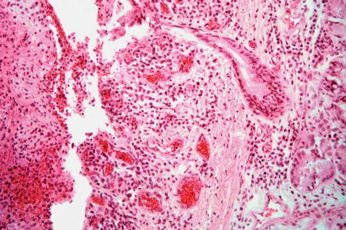
52 KB | Seung Park | This is a low-power photomicrograph of the lung from the lesion seen on x-ray. Note that there is little, if any, inflammatory reaction. | 1 |
| 04:10, 21 August 2013 | IPLab10Crypto3.jpg (file) |  |
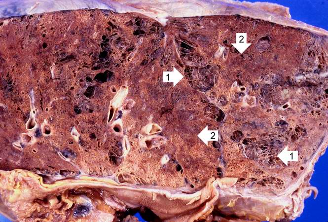
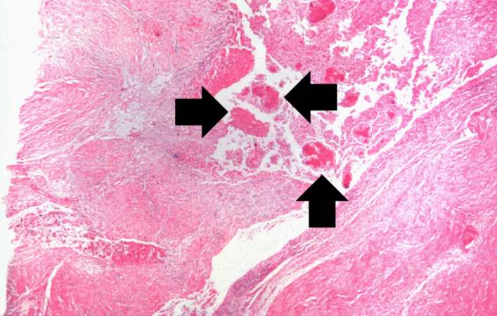
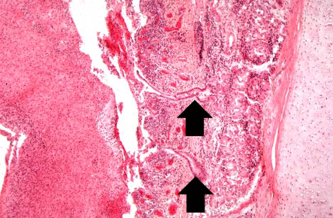
67 KB | Seung Park | This is another section of this lung showing consolidation (arrows). | 1 |
| 04:10, 21 August 2013 | IPLab10Crypto2.jpg (file) |  |
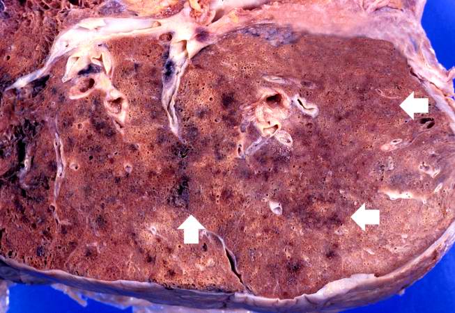
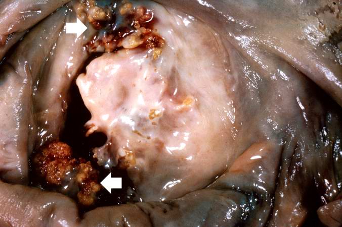
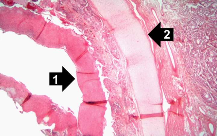
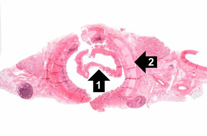
83 KB | Seung Park | This is a gross photomicrograph of this lung taken at autopsy. Note the areas of emphysema (1) and consolidation (2). | 1 |
| 04:10, 21 August 2013 | IPLab10Crypto1.jpg (file) |  |
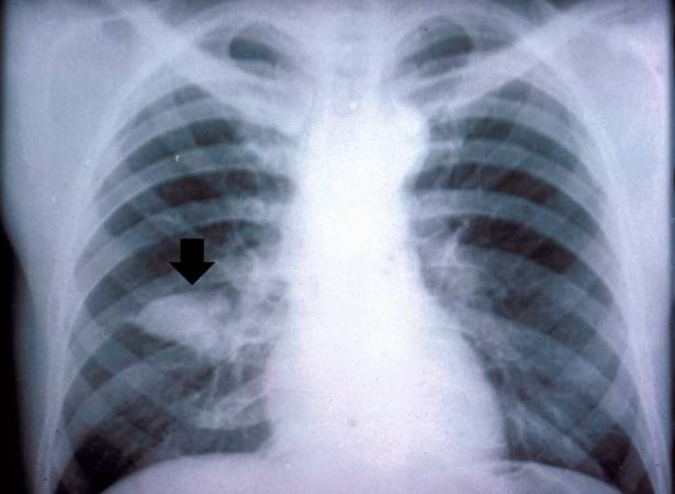
23 KB | Seung Park | This is the chest x-ray showing the mass (arrow) in the right lower lobe. | 1 |
| 04:07, 21 August 2013 | IPLab10Histo7.jpg (file) |  |
34 KB | Seung Park | This photomicrograph was taken under oil immersion to show the silver-stained Histoplasma organisms. Some of the organisms appear to be budding (arrows). | 1 |
| 04:07, 21 August 2013 | IPLab10Histo6.jpg (file) |  |
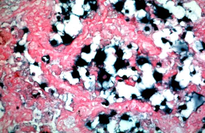
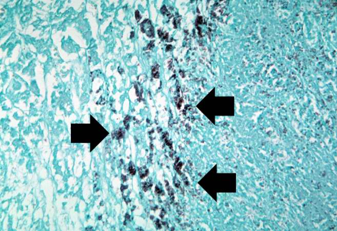
67 KB | Seung Park | This is a high-power photomicrograph of the same section of tissue as the previous slide. This section, however, has been stained with methenamine silver which causes the Histoplasma organisms to stain black (arrows). | 1 |
| 04:06, 21 August 2013 | IPLab10Histo5.jpg (file) |  |
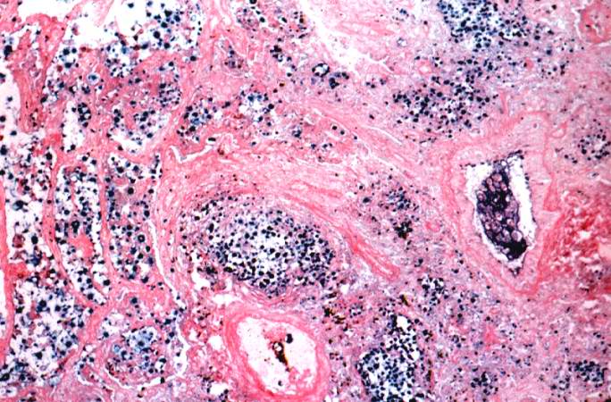
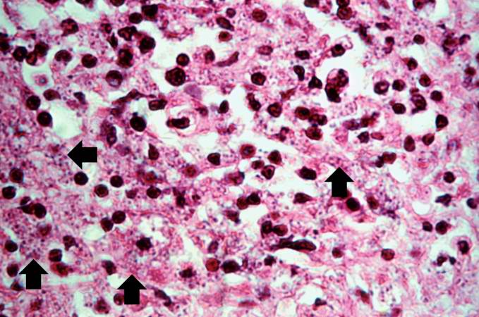
66 KB | Seung Park | This high-power photomicrograph shows small (2-5 mm) dark-staining organisms in the cytoplasm of many of these cells (arrows). | 1 |
| 04:06, 21 August 2013 | IPLab10Histo4.jpg (file) |  |
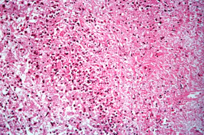
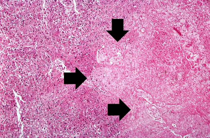
78 KB | Seung Park | This high-power photomicrograph was taken at the edge of the area of necrosis. There is a mild inflammatory infiltrate along the edge of the necrosis. | 1 |
| 04:06, 21 August 2013 | IPLab10Histo3.jpg (file) |  |
90 KB | Seung Park | This is an even higher-power photomicrograph of an area of necrosis (arrows). There is loss of cellular detail within this area. There are inflammatory cells present; however, it is difficult to differentiate the inflammatory cells from the native lymp... | 1 |
| 04:06, 21 August 2013 | IPLab10Histo2.jpg (file) |  |
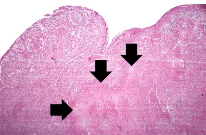
52 KB | Seung Park | This higher-power photomicrograph of the previous adrenal gland shows more clearly the irregularly-shaped area of necrosis (arrows). | 1 |
| 04:06, 21 August 2013 | IPLab10Histo1.jpg (file) |  |
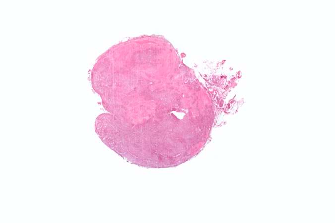
13 KB | Seung Park | This low-power photomicrograph shows a section of adrenal gland with several irregularly-outlined areas of necrosis. | 1 |
| 04:04, 21 August 2013 | IPLab10Candidiasis8.jpg (file) |  |
78 KB | Seung Park | This is a higher-power photomicrograph of a Candida colony in the kidney. Note the pseudohyphae of the Candida organisms. | 1 |
| 04:03, 21 August 2013 | IPLab10Candidiasis7.jpg (file) |  |
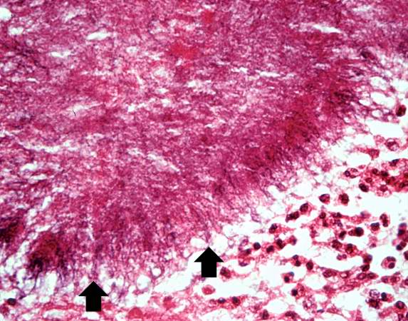
77 KB | Seung Park | This is a low-power photomicrograph of the kidney from this same case. Note the Candida colonies (arrows). The pseudohyphae are evident around the periphery of these colonies even at this low magnification. | 1 |
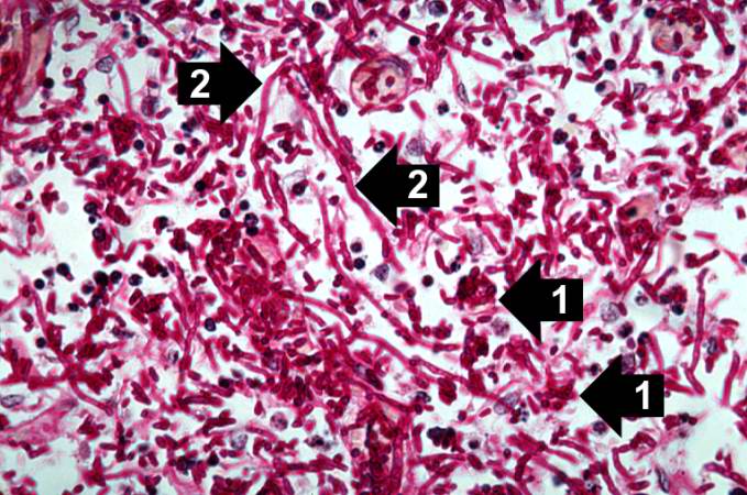
| 04:03, 21 August 2013 | IPLab10Candidiasis6.jpg (file) |  |
78 KB | Seung Park | This high-power photomicrograph shows the yeasts (1) and pseudohyphae (2). | 1 |
| 04:03, 21 August 2013 | IPLab10Candidiasis5.jpg (file) |  |
91 KB | Seung Park | This higher-power photomicrograph shows the yeasts and pseudohyphae in this focus of Candida organisms. | 1 |
| 04:03, 21 August 2013 | IPLab10Candidiasis4.jpg (file) |  |
53 KB | Seung Park | This is a low-power photomicrograph of one of the Candida colonies from this lymph node. The chains of yeast which are termed "pseudohyphae" are apparent at this magnification. | 1 |
| 04:03, 21 August 2013 | IPLab10Candidiasis3.jpg (file) |  |
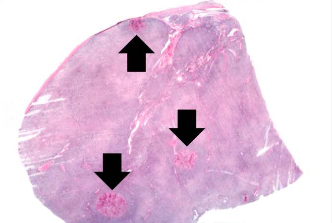
25 KB | Seung Park | This is a low-power photomicrograph of lymph node with three prominent areas of Candida colonies (arrows). Even at this low magnification, the purple-staining yeast and pseudohyphae can be easily seen. This section was stained with Periodic Acid-Schiff... | 1 |
| 04:02, 21 August 2013 | IPLab10Candidiasis2.jpg (file) |  |
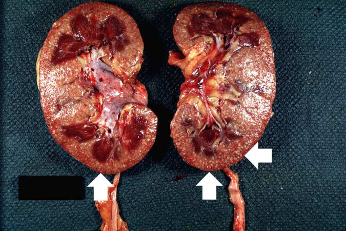
74 KB | Seung Park | This photograph of the cut surface of these kidneys shows that these multifocal punctate lesions are primarily in the cortex (arrows). | 1 |
| 04:02, 21 August 2013 | IPLab10Candidiasis1.jpg (file) |  |
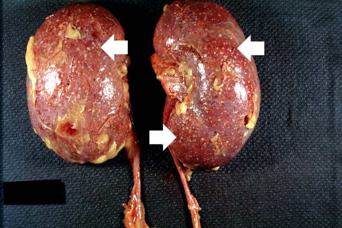
64 KB | Seung Park | This autopsy photograph of the kidneys demonstrates the multifocal punctate lesions visible on the serosal surface (arrows). Don't confuse these small yellow punctate lesions with the fat that is adherent to the renal capsule. | 1 |
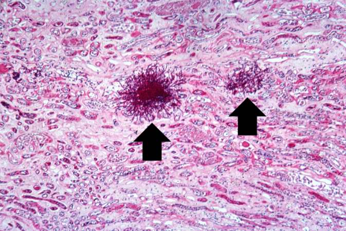
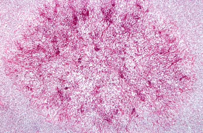
| 03:59, 21 August 2013 | IPLab9Actinomycosis5.jpg (file) |  |
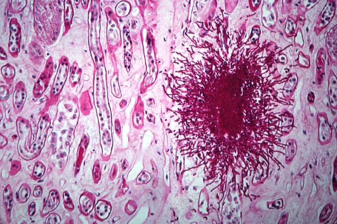
63 KB | Seung Park | This is a high-power photomicrograph of an actinomycotic colony. The filamentous nature (arrows) of the actinomyces organisms is more easily appreciated at this power. | 1 |
| 03:59, 21 August 2013 | IPLab9Actinomycosis4.jpg (file) |  |
63 KB | Seung Park | This is an even higher-power photomicrograph of actinomycotic colonies in the abscess. The filamentous nature (arrows) of the actinomyces organisms in these colonies can be appreciated. | 1 |
| 03:59, 21 August 2013 | IPLab9Actinomycosis3.jpg (file) |  |
86 KB | Seung Park | This is a higher-power photomicrograph of actinomycotic colonies in the abscess. | 1 |
| 03:59, 21 August 2013 | IPLab9Actinomycosis2.jpg (file) |  |
59 KB | Seung Park | This is a higher-power photomicrograph of an abscess demonstrating a pocket of purulent exudate that contains numerous actinomycotic colonies (arrows). | 1 |
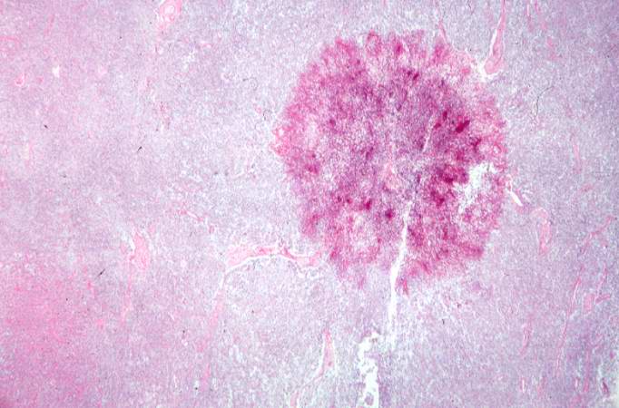
| 03:59, 21 August 2013 | IPLab9Actinomycosis1.jpg (file) |  |
39 KB | Seung Park | This is a low-power photomicrograph of the retroperitoneal abscess. At this magnification, multiple dark-staining foci can be appreciated. These foci are Actinomyces colonies (arrows). These colonies are known as "sulfur granules" because in gross spec... | 1 |
| 03:57, 21 August 2013 | IPLab9Clostridium7.jpg (file) |  |
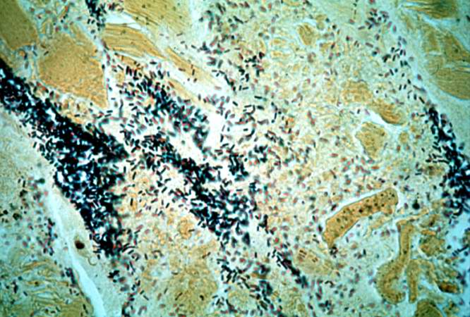
70 KB | Seung Park | This is a high-power photomicrograph of a tissue section stained with a tissue Gram's stain (Brown & Brenn). The Gram-positive bacilli can be seen throughout this tissue section. | 1 |
| 03:57, 21 August 2013 | IPLab9Clostridium6.jpg (file) |  |
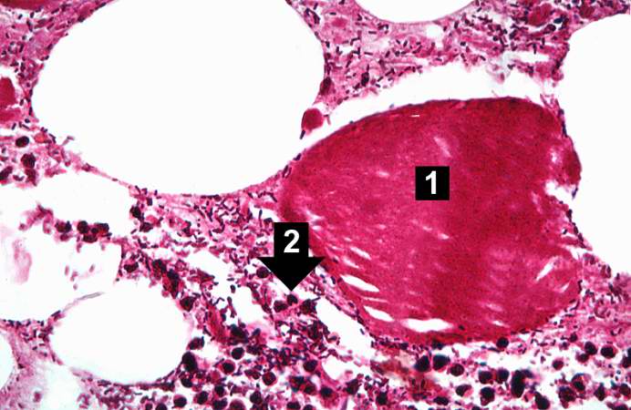
55 KB | Seung Park | This higher-power photomicrograph of the previous image provides a clearer view of gas bubbles in the tissue, the necrotic hypereosinophilic muscle cell (1), and the mild inflammatory reaction (2). At this magnification, the bacteria located throughout... | 1 |
| 03:56, 21 August 2013 | IPLab9Clostridium5.jpg (file) |  |
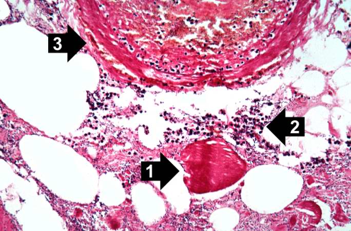
68 KB | Seung Park | This high-power photomicrograph shows the gas accumulation present in the tissue, a necrotic muscle cell (1), and a mild inflammatory response (2). There is also a thrombosed blood vessel (3). The blue-staining rods (bacterial organisms) can barely be ... | 1 |
| 03:56, 21 August 2013 | IPLab9Clostridium4.jpg (file) |  |
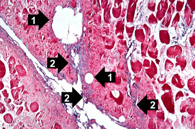
83 KB | Seung Park | This is a high-power photomicrograph of skeletal muscle. The muscle cells are hypereosinophilic and most do not contain nuclei, indicating that these cells are dead or dying. The round clear spaces (1) in this tissue correspond to gas accumulations pri... | 1 |
| 03:56, 21 August 2013 | IPLab9Clostridium3.jpg (file) |  |
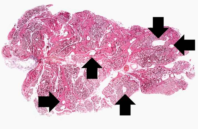
62 KB | Seung Park | This is a low-power photomicrograph of muscle fascicles containing large gas bubbles (arrows). Note that there is no inflammatory reaction in this section. | 1 |
| 03:56, 21 August 2013 | IPLab9Clostridium2.jpg (file) |  |
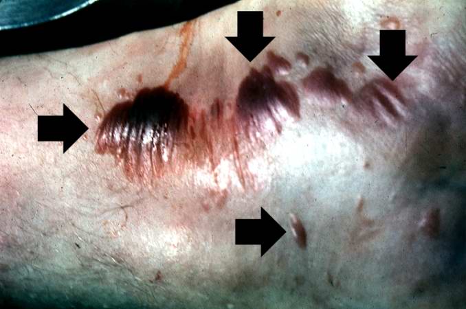
40 KB | Seung Park | This gross photograph shows a close-up view of hemorrhagic blebs (arrows) on the skin. The blebs on the skin are accumulations of gas being discharged into the tissues from the Clostridium perfringens. This gas produces crepitance. | 1 |
| 03:56, 21 August 2013 | IPLab9Clostridium1.jpg (file) |  |
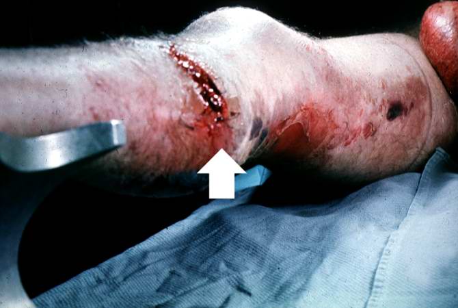
36 KB | Seung Park | This gross photograph of the lower extremity was taken at autopsy. Notice the swelling and the area of the primary infection (arrow). | 1 |
| 03:53, 21 August 2013 | IPLab9ARF6.jpg (file) |  |
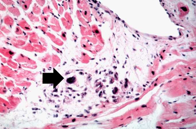
50 KB | Seung Park | This high-power photomicrograph of myocardium shows the cellular detail of another Aschoff body. In this case there appears to be a multinucleated Aschoff giant cell (arrow). | 1 |
| 03:52, 21 August 2013 | IPLab9ARF5.jpg (file) |  |
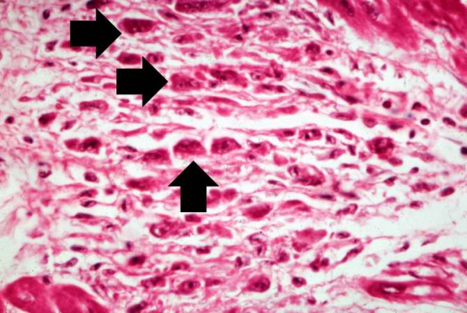
52 KB | Seung Park | This high-power photomicrograph of myocardium shows the cellular detail of an Aschoff body. Aschoff bodies are foci of fibrinoid necrosis surrounded by lymphocytes, macrophages, an occasional plasma cell, and plump “activated” histiocytes called An... | 1 |
| 03:52, 21 August 2013 | IPLab9ARF4.jpg (file) |  |
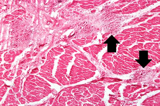
76 KB | Seung Park | This is a higher-power photomicrograph of myocardium containing Aschoff bodies (arrows) within the interstitium. | 1 |
| 03:52, 21 August 2013 | IPLab9ARF3.jpg (file) |  |
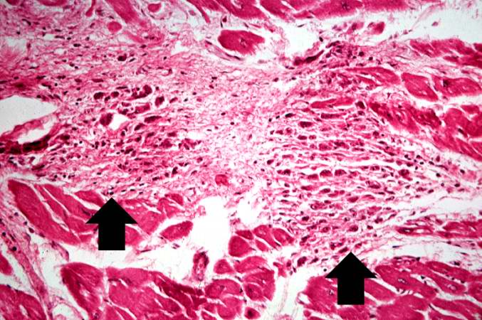
83 KB | Seung Park | This is a higher-power photomicrograph of myocardium showing cellular accumulations--Aschoff bodies (arrows)--within the interstitium of the myocardium. These are found especially around blood vessels. | 1 |
| 03:52, 21 August 2013 | IPLab9ARF2.jpg (file) |  |
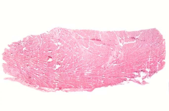
29 KB | Seung Park | This is a low-power photomicrograph of heart tissue. Little can be seen at this magnification, except that the tissue looks relatively normal. | 1 |
| 03:52, 21 August 2013 | IPLab9ARF1.jpg (file) |  |
48 KB | Seung Park | This is a gross photograph of mitral valve demonstrating marked thickening and fibrosis of the valve leaflet. There are also numerous foci of fibrinoid necrosis within the cusps and friable vegetations (verrucae) along the lines of closure (arrows). Th... | 1 |
| 03:50, 21 August 2013 | IPLab9Diphtheria4.jpg (file) |  |
69 KB | Seung Park | In this higher-power photomicrograph of the tissue from the previous image, the ulcerated tracheal mucosa and the diphtheritic membrane are more clearly seen. Although difficult to make out at this magnification, most of the cells in this inflammatory ... | 1 |
| 03:50, 21 August 2013 | IPLab9Diphtheria3.jpg (file) |  |
71 KB | Seung Park | This is an even higher-power photomicrograph of the tracheal mucosa and the diphtheritic membrane. The mucosal surface of the trachea is ulcerated (total loss of epithelial cells) and the only remaining epithelial cells are found in the glands (arrows)... | 1 |
| 03:49, 21 August 2013 | IPLab9Diphtheria2.jpg (file) |  |
47 KB | Seung Park | This is a higher-power photomicrograph of trachea with the diphtheritic membrane (1). Even though, the main part of the membrane has pulled away from the tracheal lining during histological processing, in this section part of the membrane is still loos... | 1 |
| 03:49, 21 August 2013 | IPLab9Diphtheria1.jpg (file) |  |
28 KB | Seung Park | This is a low-power photomicrograph of the trachea with the diphtheritic membrane (1), which has pulled away from the tracheal lining during histological processing. Note the tracheal cartilage (2) present in this section. | 1 |
| 03:47, 21 August 2013 | IPLab9BacterialMeningitis8.jpg (file) |  |
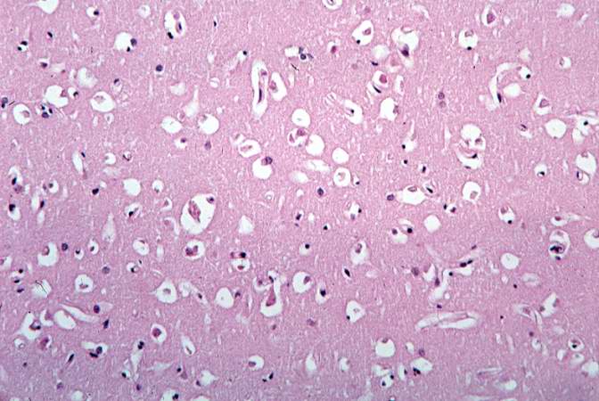
56 KB | Seung Park | This photomicrograph of brain tissue demonstrates diffuse edema. | 1 |