IPLab:Lab 6:Graves Disease: Difference between revisions
Jump to navigation
Jump to search
Seung Park (talk | contribs) Created page with "== Images == <gallery heights="250px" widths="250px"> File:IPLab6GravesDisease1.jpg|This is a photograph of the thyroid from this case. Note that the gland is enlarged and is ..." |
(No difference)
|
Revision as of 13:23, 19 August 2013
Images
-
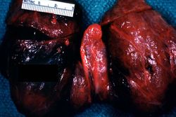 This is a photograph of the thyroid from this case. Note that the gland is enlarged and is dark red.
 -
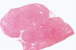 This is a low-power photomicrograph of thyroid tissue from this case. The tissue is very cellular with very little colloid.
 -
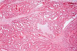 This is a higher-power photomicrograph of thyroid. The tissue is very cellular and there is little colloid.
 -
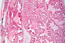 This is a high-power photomicrograph of thyroid. Note the cellularity of the tissue with marked infolding of the epithelial tissue.
 -
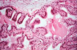 This is a high-power photomicrograph of thyroid. Note the papillary projections and the moth-eaten appearance of the colloid. This appearance indicates active absorption of the colloid to form thyroglobulin.
 -
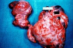 This is a gross photograph of a thyroid from a case of nodular goiter.
 -
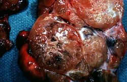 Closer view of cut surface of the thyroid with nodular goiter. Note the multilobular appearance of the tissue.


