Difference between revisions of "IPLab:Lab 5:Neurofibromatosis"
| Line 12: | Line 12: | ||
</gallery> | </gallery> | ||
| − | {{IPLab | + | {{IPLab 5}} |
[[Category: IPLab:Lab 5]] | [[Category: IPLab:Lab 5]] | ||
Revision as of 17:30, 19 August 2013
Images
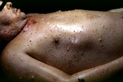
This photograph, taken at autopsy, demonstrates the distribution of neurofibromas on the skin of this patient.
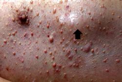
This is another view taken at autopsy demonstrating the neurofibromas. Some lesions can be seen as subcutaneous swellings (arrow) and others form pedunculated masses. Most are hyperpigmented.
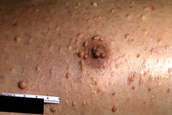
This is a closer view of neurofibromas on the skin.
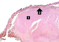
This is a low-power photomicrograph of a subcutaneous neurofibroma (1). Note the increased pigmentation in the skin (2).
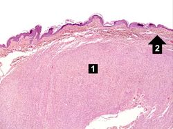
This is a higher-power photomicrograph of the neurofibroma (1) with the overlying skin (2).
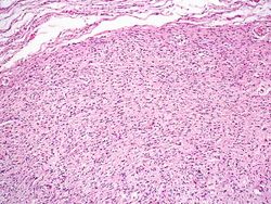
This is a higher-power photomicrograph of the neurofibroma demonstrating the loose pattern of elongated cells making up the tumor mass.
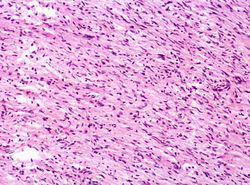
This higher-power photomicrograph of the neurofibroma shows more clearly the elongated cells (primarily Schwann cells) that make up this tumor.
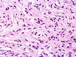
This is a high-power photomicrograph of the cells in the neurofibroma.


| |||||