Cytologically Yours: CoW: 20131202: Difference between revisions
Jump to navigation
Jump to search
No edit summary |
No edit summary |
||
| Line 38: | Line 38: | ||
====Resident Questions==== | ====Resident Questions==== | ||
* <spoiler text="What is your differential diagnosis?">These groups of cells demonstrate malignant appearing cells in a background of an otherwise benign appearing lymphoid background. The atypical cells are scattered, with large nucleoli and several binucleate forms. In addition, there seem to be an increased number of eosinophils in the background. The differential diagnosis includes Hodgkin lymphoma; however, the possibility of the large atypical cells being melanoma cannot be ruled out. </spoiler> | * <spoiler text="What is your differential diagnosis?">These groups of cells demonstrate malignant appearing cells in a background of an otherwise benign appearing lymphoid background. The atypical cells are scattered, with large nucleoli and several binucleate forms. In addition, there seem to be an increased number of eosinophils in the background. The differential diagnosis includes Hodgkin lymphoma; however, the possibility of the large atypical cells being melanoma cannot be ruled out. </spoiler> | ||
* <spoiler text="What would you | * <spoiler text="What ancillary tests would you recommend?">For this patient, we recommended that the radiologist perform a biopsy of the lesion so that it could be sent for immunohistochemical workup. Since the overall percentage of the atypical cells were low, we were worried that a cell block would not contain enough of the malignant cells for additional stains. We also sent the lymph node for flow since a hematologic malignancy was suspected; however, with Hodgkin lymphoma, we don't expect any diagnostic findings from flow cytometry.</spoiler> | ||
===Biopsy=== | ===Biopsy=== | ||
| Line 67: | Line 67: | ||
==Final Diagnosis== | ==Final Diagnosis== | ||
===Cytology=== | ===Cytology=== | ||
* '''Positive for malignancy'''. | * '''Positive for malignancy, the differential diagnosis includes melanoma and Hodgkin lymphoma'''. | ||
===Biopsy=== | ===Biopsy=== | ||
* ''' | * '''Classical Hodgkin lymphoma, favor mixed type'''. | ||
==Case Discussion== | ==Case Discussion== | ||
Revision as of 15:09, 14 January 2014
Clinical Summary
The patient is an 60 year old male with a remote history of an abdominal melanoma that was excised with negative margins. The patient has been experiencing lower back pain for the past several months and has received epidural injections. As a part of the workup, the patient had a CT which revealed retroperitoneal lymphadenopathy. A CT guided fine needle aspiration and biopsy of a paracaval lymph node was performed.
Past Medical History
- 2003 Melanoma
- Diabetes
- Hypertension
Past Surgical History
- 2013 Arthroscopic knee surgery
- 2003 Excision of melanoma
- 2002 Discectomy
Clinical Plan
The differential diagnosis for otherwise asymptomatic lymphadenopathy in this patient is melanoma, lymphoma, or occult malignancy.
Radiology
- PET CT showed hypermetabolic activity with an SUV of 12.7.
- CT of abdomen and pelvis showed adenopathy adjacent to the aorta and inferior to the vena cava at the level of the right kidney. The largest node measured 4 cm in greatest dimension.
CT


Pathology
Cytology
-
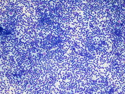 10x magnification of paracaval lymph node. There is a polymorphic lymphoid population with scattered large atypical cells.
 -
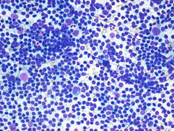 20x magnification of paracaval lymph node. There are small lymphocytes with background lymphoglandular bodies. Scattered eosinophils and large atypical cells with prominent nucleoli.
 -
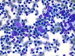 40x magnification of paracaval lymph node. There are atypical binucleated cells among the large atypical cells.


Resident Questions
Biopsy
-
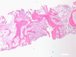
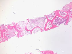 This is a 2x magnification the biopsy specimen from the spinal mass. We see that this is bone histology which is abnormal. There is an increase in cellularity, especially for an 80 year old person.
 -
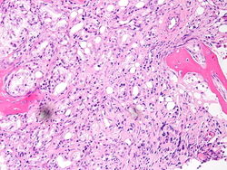
 This is a 4x magnification of the biopsy specimen from the spinal mass. We can see that there is an increase in cellularity.
 -
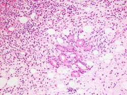
 This is a 20x magnification of the biopsy specimen from the spinal mass. This is one of the areas that has increased cellularity on lower power. We can see cells forming what appear to be glands in the bone marrow.
 -
 This is a 20x magnification of the biopsy specimen from the spinal mass. Here we see well formed glands in the bone marrow.
 -
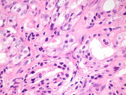 This is a 60x magnification of the biopsy specimen from the spinal mass. The malignant cells appear to be creating glandular structures, and we can identify prominent nucleoli in the malignant cells.
 -
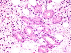 This is a 60x magnification of the biopsy specimen from the spinal mass. Again we see well formed glands in the bone marrow, made of cells that have prominent nucleoli.


Immunohistochemistry
-
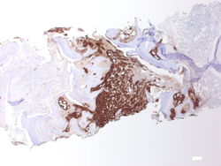 PSA, 2x magnification. Note the predominance of brown staining in the middle.
 -
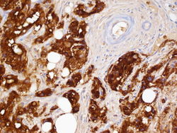 PSA, 60x magnification. The glands are highlighted in brown, and on the upper right the blood vessels are negative.


Resident Questions
Final Diagnosis
Cytology
- Positive for malignancy, the differential diagnosis includes melanoma and Hodgkin lymphoma.
Biopsy
- Classical Hodgkin lymphoma, favor mixed type.
Case Discussion
This is a classic case of prostatic adenocarcinoma, metastatic to the spine.
| ||||||||