IPLab:Lab 11:Chagas Disease: Difference between revisions
Jump to navigation
Jump to search
Seung Park (talk | contribs) No edit summary |
Seung Park (talk | contribs) |
||
| Line 18: | Line 18: | ||
== Additional Resources == | == Additional Resources == | ||
=== Reference === | === Reference === | ||
* [http://emedicine.medscape.com/article/214581-overview eMedicine Medical Library: Chagas Disease (American Trypanosomiasis)] | |||
* [http://emedicine.medscape.com/article/1000389-overview eMedicine Medical Library: Trypanosomiasis] | |||
* [http://www.merckmanuals.com/professional/infectious_diseases/extraintestinal_protozoa/chagas_disease.html Merck Manual: Chagas Disease] | |||
* [http://www.merckmanuals.com/professional/infectious_diseases/extraintestinal_protozoa/african_trypanosomiasis.html Merck Manual: African Trypanosomiasis] | |||
=== Journal Articles === | === Journal Articles === | ||
Revision as of 23:49, 24 August 2013
Clinical Summary
This 12-year-old boy, whose family had recently emigrated from Brazil, presented to the emergency room with a three-day history of malaise, fever, anorexia, and edema of the face and upper extremities. On physical examination the patient had generalized lymphadenopathy and hepatosplenomegaly. The patient was tachycardic and dysgenic with signs of congestive heart failure. A cardiac biopsy was performed which revealed an active myocarditis with leishmanial forms of parasitic organisms within cardiac myocytes. Close examination of peripheral blood smears revealed occasional circulating trypomastigotes. A complement fixation test for antibodies to Trypanosoma cruzi was strongly positive.
Images
-
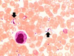 This peripheral blood smear from the patient shows two trypomastigotes of Trypanosoma cruzi.
 -
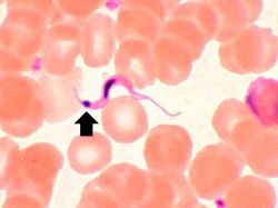 This peripheral blood smear from the patient shows a higher power view of a Trypanosoma cruzi trypomastigote. Note the prominent kinetoplast (arrow).
 -
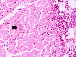 This is a low-power photomicrograph of an H & E stained section from the heart biopsy of this patient. Note the organisms within a myocyte (arrow) and the adjacent inflammatory response.
 -
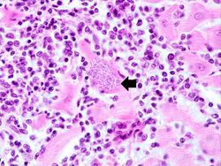 This is a higher-power photomicrograph of an H & E stained heart biopsy from this patient. Again, note the organisms within a myocyte (arrow) and the inflammatory response.
 -
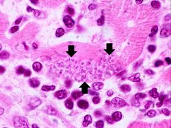 This is a higher-power photomicrograph of an H & E stained heart biopsy from this patient. At this magnification the organisms within a myocyte (arrows) and the adjacent inflammatory response are more clearly seen. The individual organisms within the myocyte are called amastigotes.
 -
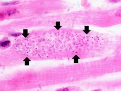 This is a higher-power photomicrograph of an H & E stained heart biopsy from this patient. Note the T. cruzi amastigotes (arrows) within this longitudinal section of a myocyte.


Study Questions
Additional Resources
Reference
- eMedicine Medical Library: Chagas Disease (American Trypanosomiasis)
- eMedicine Medical Library: Trypanosomiasis
- Merck Manual: Chagas Disease
- Merck Manual: African Trypanosomiasis
Journal Articles
Images
Related IPLab Cases
| |||||