Difference between revisions of "File:IPLab1Tuberculosis7.jpg"
Seung Park (talk | contribs) |
Seung Park (talk | contribs) |
||
| Line 1: | Line 1: | ||
| − | + | This is a higher-power view of the granuloma with the amorphous eosinophilic material representing caseation necrosis (1), giant cells near the center (2), and inflammatory cells around the periphery. | |
Latest revision as of 02:53, 16 August 2013
This is a higher-power view of the granuloma with the amorphous eosinophilic material representing caseation necrosis (1), giant cells near the center (2), and inflammatory cells around the periphery.
File history
Click on a date/time to view the file as it appeared at that time.
| Date/Time | Thumbnail | Dimensions | User | Comment | |
|---|---|---|---|---|---|
| current | 02:50, 16 August 2013 | 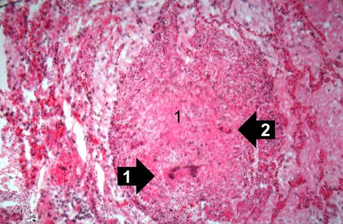 | 687 × 450 (64 KB) | Seung Park (talk | contribs) |
- You cannot overwrite this file.
File usage
There are no pages that link to this file.