File:IPLab1KidneyInfarction4.jpg: Difference between revisions
Jump to navigation
Jump to search

Seung Park (talk | contribs) No edit summary |
Seung Park (talk | contribs) No edit summary |
||
| Line 1: | Line 1: | ||
A higher-power photomicrograph shows the edge of this reddish area, illustrating coagulation necrosis (1) compared to the normal tissue (2). The necrotic tubules in this hemorrhagic, red infarct are hypereosinophilic. Compare the tubules on the right with the normal tubules seen in the left-hand portion of the slide. Note the interstitial hemorrhage which is associated with vascular leakage within this necrotic region in the tissue. | |||


Revision as of 11:18, 15 August 2013
A higher-power photomicrograph shows the edge of this reddish area, illustrating coagulation necrosis (1) compared to the normal tissue (2). The necrotic tubules in this hemorrhagic, red infarct are hypereosinophilic. Compare the tubules on the right with the normal tubules seen in the left-hand portion of the slide. Note the interstitial hemorrhage which is associated with vascular leakage within this necrotic region in the tissue.
File history
Click on a date/time to view the file as it appeared at that time.
| Date/Time | Thumbnail | Dimensions | User | Comment | |
|---|---|---|---|---|---|
| current | 16:15, 27 June 2019 | 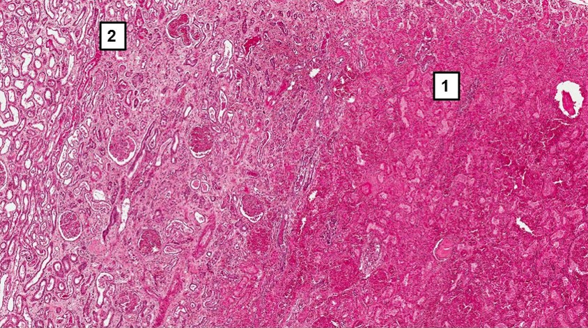 | 844 × 472 (228 KB) | Peter Anderson (talk | contribs) | |
| 10:12, 15 August 2013 | 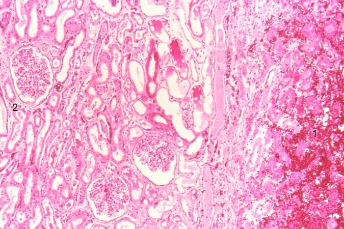 | 675 × 450 (71 KB) | Seung Park (talk | contribs) |
You cannot overwrite this file.
File usage
The following page uses this file:
