Difference between revisions of "IPLab:Lab 11:Malaria"
Seung Park (talk | contribs) (Created page with "== Images == <gallery heights="250px" widths="250px"> File:IPLab11Malaria1.jpg|This is a high power photomicrograph of a thin smear of blood from this patient. Note that one o...") |
(No difference)
|
Revision as of 04:55, 21 August 2013
Images
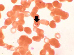
This is a high power photomicrograph of a thin smear of blood from this patient. Note that one of the RBCs has a ring stage trophozoite (arrow).
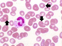
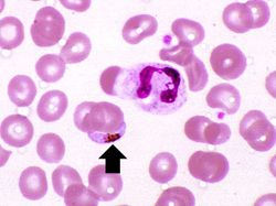
This is another high power photomicrograph of a thin smear of blood from this patient. There is a single eosinophil in this smear along with several RBCs containing ring stage trophozoites (arrows).
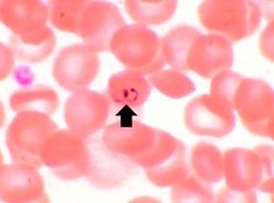
This is yet another high power photomicrograph of a thin smear of blood from this patient. There is one RBC that contains two ring stage trophozoites (arrow). This is characteristic of, but not diagnostic for, P. falciparum.
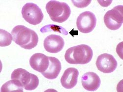
In this high power photomicrograph of a thin smear of blood from this patient there is one P. falciparum gametocyte (arrow). These gametocytes have a characteristic "banana" shape.
There is another example of a P. falciparum gametocyte (arrow) in this thin smear. There is a neutrophil in this field as well.
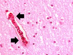
This high photomicrograph was taken from another patient who died of malignant cerebral malaria caused by P. falciparum. In this photomicrograph, a small artery (arrow) can be seen that is full of parasitized RBCs. These RBCs tend to clog small blood vessels and lead to cerebral ischemia/hypoxia.
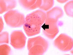
In this peripheral smear from a different patient who was infected with P. vivax, the cytoplasm of the infected RBC has a stippled appearance (Schüffner's dots) (arrow). The RBC is also slightly enlarged.


| |||||