IPLab:Lab 9:RMSF: Difference between revisions
Jump to navigation
Jump to search
Seung Park (talk | contribs) Created page with "== Images == <gallery heights="250px" widths="250px"> File:IPLab9RMSF1.jpg|This is a low-power photomicrograph of the skin biopsy. Note areas of hemorrhage (arrow) in the derm..." |
Seung Park (talk | contribs) No edit summary |
||
| Line 1: | Line 1: | ||
== Clinical Summary == | |||
This 9-year-old child was admitted with headache, fever, and a morbilliform rash on the arms and legs. There was a history of a tick being removed from her back. By the time a biopsy was performed, the rash had become petechial. Antibiotics were given and the child recovered within one week. | |||
== Biopsy Findings == | |||
A skin biopsy of this patient's lesion was stained with hematoxylin and eosin. A different section was also stained with an immunoperoxidase technique using antibody against Rickettsia rickettsii. Organisms were demonstrated in the endothelial cells. | |||
== Images == | == Images == | ||
<gallery heights="250px" widths="250px"> | <gallery heights="250px" widths="250px"> | ||
Revision as of 09:36, 21 August 2013
Clinical Summary
This 9-year-old child was admitted with headache, fever, and a morbilliform rash on the arms and legs. There was a history of a tick being removed from her back. By the time a biopsy was performed, the rash had become petechial. Antibiotics were given and the child recovered within one week.
Biopsy Findings
A skin biopsy of this patient's lesion was stained with hematoxylin and eosin. A different section was also stained with an immunoperoxidase technique using antibody against Rickettsia rickettsii. Organisms were demonstrated in the endothelial cells.
Images
-
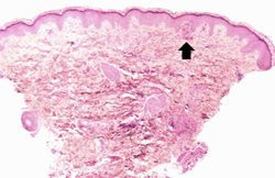 This is a low-power photomicrograph of the skin biopsy. Note areas of hemorrhage (arrow) in the dermis.
 -
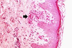 This is a higher-power photomicrograph demonstrating areas of hemorrhage immediately underneath the epidermis. Also note the cellularity and thrombosis of the small vessels in the dermis (arrow).
 -
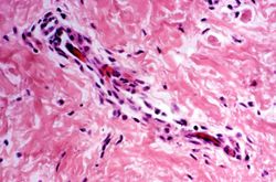 This is a photomicrograph of a small vessel in the dermis which is demonstrating mild vasculitis.
 -
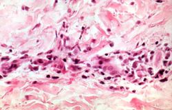 This is a higher-power photomicrograph of a dermal vessel with mild vasculitis.
 -
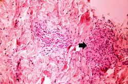 This is a photomicrograph of dermis with an area of more severe vasculitis (arrow).
 -
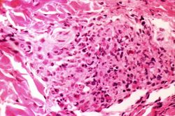 This is a high-power photomicrograph of severe vasculitis in the dermis.
 -
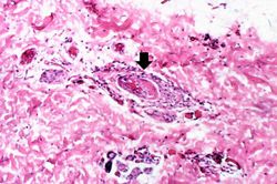 This is a high-power photomicrograph of a dermal vessel (arrow) which is exhibiting vasculitis and thrombosis.
 -
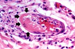 This is a high-power photomicrograph of a thrombosed vessel in the dermis. Note that the endothelial cells are missing along part of the circumference of the vessel (arrows)--this is where the main part of the thrombus has attached. Also note the inflammation surrounding the vessel.


| |||||