IPLab:Lab 6:Graves Disease: Difference between revisions
Jump to navigation
Jump to search
Seung Park (talk | contribs) Created page with "== Images == <gallery heights="250px" widths="250px"> File:IPLab6GravesDisease1.jpg|This is a photograph of the thyroid from this case. Note that the gland is enlarged and is ..." |
Seung Park (talk | contribs) No edit summary |
||
| Line 1: | Line 1: | ||
== Clinical Summary == | |||
This 18-year-old girl presented with complaints of swelling in the neck, weight loss, bulging of the eyes, tremor, decreased heat tolerance, loose stools, and occasional palpitations. Physical examination revealed normal blood pressure, resting tachycardia of 110 beats per minute, mild exophthalmos, eyelid lag, and a diffusely enlarged thyroid gland. Pertinent laboratory findings were thyroxine (T4) level 30.8 mcg/dL, free thyroxine was 2.7 ng/dL, and thyroid stimulating hormone (TSH) was 0.22 mcIU/mL. She was given propylthiouracil until she became nearly euthyroid, at which time a thyroidectomy was done. | |||
== Autopsy Findings == | |||
The thyroid gland weighed 45 grams. It was beefy red in color and had a homogeneous fleshy consistency. | |||
== Images == | == Images == | ||
<gallery heights="250px" widths="250px"> | <gallery heights="250px" widths="250px"> | ||
Revision as of 09:23, 21 August 2013
Clinical Summary
This 18-year-old girl presented with complaints of swelling in the neck, weight loss, bulging of the eyes, tremor, decreased heat tolerance, loose stools, and occasional palpitations. Physical examination revealed normal blood pressure, resting tachycardia of 110 beats per minute, mild exophthalmos, eyelid lag, and a diffusely enlarged thyroid gland. Pertinent laboratory findings were thyroxine (T4) level 30.8 mcg/dL, free thyroxine was 2.7 ng/dL, and thyroid stimulating hormone (TSH) was 0.22 mcIU/mL. She was given propylthiouracil until she became nearly euthyroid, at which time a thyroidectomy was done.
Autopsy Findings
The thyroid gland weighed 45 grams. It was beefy red in color and had a homogeneous fleshy consistency.
Images
-
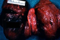 This is a photograph of the thyroid from this case. Note that the gland is enlarged and is dark red.
 -
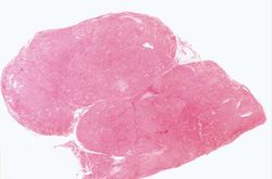 This is a low-power photomicrograph of thyroid tissue from this case. The tissue is very cellular with very little colloid.
 -
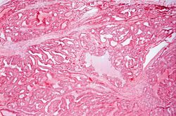 This is a higher-power photomicrograph of thyroid. The tissue is very cellular and there is little colloid.
 -
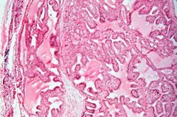 This is a high-power photomicrograph of thyroid. Note the cellularity of the tissue with marked infolding of the epithelial tissue.
 -
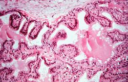 This is a high-power photomicrograph of thyroid. Note the papillary projections and the moth-eaten appearance of the colloid. This appearance indicates active absorption of the colloid to form thyroglobulin.
 -
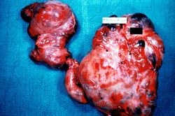 This is a gross photograph of a thyroid from a case of nodular goiter.
 -
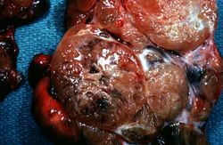 Closer view of cut surface of the thyroid with nodular goiter. Note the multilobular appearance of the tissue.


