IPLab:Lab 1:Fat Necrosis: Difference between revisions
Jump to navigation
Jump to search
| Line 13: | Line 13: | ||
File:IPLab1FatNecrosis6.jpg|A higher-power photomicrograph of the previous slide contains a small area of fat necrosis (1) in the upper right portion of the image. The fat necrosis is within the fat tissue that is normally found adjacent to the pancreas. The appearance of the pancreatic tissue in this area is somewhat disrupted due to autolysis (the pancreas autolyzes very rapidly after death) but there is some premortem necrosis as well. | File:IPLab1FatNecrosis6.jpg|A higher-power photomicrograph of the previous slide contains a small area of fat necrosis (1) in the upper right portion of the image. The fat necrosis is within the fat tissue that is normally found adjacent to the pancreas. The appearance of the pancreatic tissue in this area is somewhat disrupted due to autolysis (the pancreas autolyzes very rapidly after death) but there is some premortem necrosis as well. | ||
File:IPLab1FatNecrosis7.jpg|This is a higher-power photomicrograph of the fat necrosis involving the fat cells in the interlobular spaces (arrow) of the pancreas. Note the blue to purple staining of the calcium deposits within the fat cells. | File:IPLab1FatNecrosis7.jpg|This is a higher-power photomicrograph of the fat necrosis involving the fat cells in the interlobular spaces (arrow) of the pancreas. Note the blue to purple staining of the calcium deposits within the fat cells. | ||
File: | File:IPLab1FatNecrosis10.jpg|This high-power photomicrograph demonstrates fat necrosis in the interlobular spaces of the pancreas. Note the granular blue-staining calcium deposits (arrows) within the fat cells. The clear areas represent artifact caused by the "washing-out" of fat from cells during tissue processing for histology. | ||
</gallery> | </gallery> | ||
Revision as of 16:59, 27 June 2019
Clinical Summary
This was a 37-year-old female with chronic renal failure who underwent a cadaveric renal transplant. Following transplantation she developed oral candidiasis, pneumonia, pyuria, and gastrointestinal bleeding. Subsequently, the patient became septic and died.
Major findings at autopsy included extensive hemorrhagic bronchopneumonia and multiple ulcers affecting the stomach and esophagus. There was also evidence of disseminated intravascular coagulation (DIC) with multiple hemorrhages present. Firm, whitish foci of necrotic tissue were found in the fat around the pancreas.
Images
-
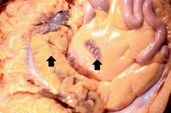 This gross photograph shows the intestines and omentum at autopsy. Note the small (5-15 mm in diameter) white nodules on the surface of the omental and mesenteric fat tissue (arrows).
 -
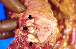 This gross photograph of the pancreas from this case shows white nodules (arrows) in the pancreas and the adjacent mesenteric fat tissue.
 -
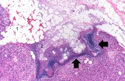 This low-power photomicrograph of the pancreas from this case shows the fat tissue (1) surrounding the pancreas. Note the rim of inflammatory cells (arrows) and the blue areas in the fat adjacent to the pancreas (2).
 -
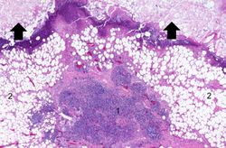 This high-power photomicrograph shows areas of inflammation (1) and fat necrosis (arrows) in the peripancreatic fat tissue (2) of the pancreas from this case.
 -
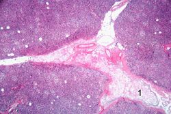 Another high-power photomicrograph shows blue discoloration in the fat tissue in the interlobular spaces (1) of the pancreas.
 -
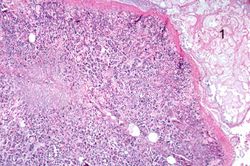 A higher-power photomicrograph of the previous slide contains a small area of fat necrosis (1) in the upper right portion of the image. The fat necrosis is within the fat tissue that is normally found adjacent to the pancreas. The appearance of the pancreatic tissue in this area is somewhat disrupted due to autolysis (the pancreas autolyzes very rapidly after death) but there is some premortem necrosis as well.
 -
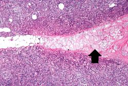 This is a higher-power photomicrograph of the fat necrosis involving the fat cells in the interlobular spaces (arrow) of the pancreas. Note the blue to purple staining of the calcium deposits within the fat cells.
 -
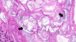 This high-power photomicrograph demonstrates fat necrosis in the interlobular spaces of the pancreas. Note the granular blue-staining calcium deposits (arrows) within the fat cells. The clear areas represent artifact caused by the "washing-out" of fat from cells during tissue processing for histology.


Virtual Microscopy
Pancreatic Fat Necrosis
Normal Pancreas
Study Questions
Additional Resources
Reference
Journal Articles
- Forsmark CE, Vege SS, Wilcox CM. Acute Pericarditis. N Engl J Med" 2016; 375:1972-1981. November 17, 2016.
- LeWinter MM. Acute Pericarditis. NEJM 2014 Dec 18;371:2410-2416.
Images
| |||||