Cytologically Yours: CoW: 20131216: Difference between revisions
Jump to navigation
Jump to search
No edit summary |
|||
| Line 56: | Line 56: | ||
<div class="usermessage mw-customtoggle-diagnosis" style="cursor:pointer">Click here to toggle the diagnosis | <div class="usermessage mw-customtoggle-diagnosis" style="cursor:pointer">Click here to toggle the diagnosis.</div> | ||
<div class="mw-collapsible mw-collapsed" id="mw-customcollapsible-diagnosis"> | <div class="mw-collapsible mw-collapsed" id="mw-customcollapsible-diagnosis"> | ||
<div class="mw-collapsible-content"> | <div class="mw-collapsible-content"> | ||
| Line 64: | Line 64: | ||
* Small cell carcinoma | * Small cell carcinoma | ||
</div></div> | </div></div> | ||
Revision as of 15:48, 15 January 2014
Clinical Summary
The patient is an 66 year old white male with a history of smoking, COPD, and diabetes. The patient presented with increased shortness of breath.
Past Medical History
- Diabetes
- COPD
- Squamous cell carcinoma of skin
Past Surgical History
- Excision of squamous cell carcinoma
- Removal of adenomatous polyp of sigmoid colon
Clinical Plan
The differential diagnosis includes worsening of COPD. CT imaging of chest is performed.
Radiology
- CT Chest shows hilar lung mass and multiple mediastinal lymph nodes showing increased uptake on PET scan.
Pathology
Cytology
-
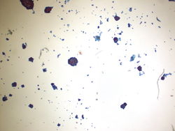 10x magnification of pleural fluid(ThinPrep). Groups of cohesive epithelial appearing cells are seen on low power.
 -
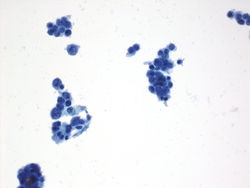 40x magnification of pleural fluid (ThinPrep). Cluster of atypical cells showing nuclear pleomorphism and scant cytoplasm.
 -
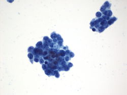 40x magnification of pleural fluid (ThinPrep). Chromatin is irregular and clumped with salt and pepper appearance; although, occasional nucleoli are also seen.
 -
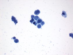 40x magnification of pleural fluid (ThinPrep). Some nuclear molding can be appreciated and a mitotic figure is present.
 -
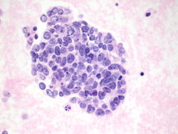 Cell block of pleural fluid. Group of malignant cells showing nuclear molding, scant cytoplasm, and salt and pepper chromatin. Nucleoli are also seen.


Immunohistochemistry
-
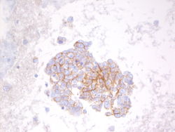 CD56 on pleural fluid shows positive cytoplasmic staining.
 -
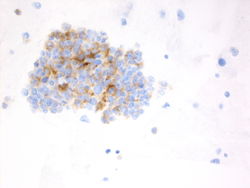 Synaptophysin on pleural fluid shows positive cytoplasmic staining.


Other immunostains performed
- BerEp4 Positive
- Moc31 Faintly positive
- Calretinin Negative
- TTF1 Negative
- Chromogranin Positive
- Synaptophysin Positive
- CD56 Positive
- Napsin A Negative
Resident Questions
Final Diagnosis
Cytology
- Small cell carcinoma
| ||||||||