IPLab:Lab 1:Kidney Infarction: Difference between revisions
Jump to navigation
Jump to search
Seung Park (talk | contribs) No edit summary |
Seung Park (talk | contribs) No edit summary |
||
| Line 6: | Line 6: | ||
== Images == | == Images == | ||
<gallery> | <gallery heights="250px" widths="250px"> | ||
File:IPLab1KidneyInfarction1.jpg|This gross photograph shows a kidney that has been transected longitudinally at autopsy. The cut surface (right) shows several areas of infarction. The most recent infarct is seen at the top left (arrow). The surface of the kidney (left) shows a marked nodularity and roughening from scarring due to chronic hypertension. | File:IPLab1KidneyInfarction1.jpg|This gross photograph shows a kidney that has been transected longitudinally at autopsy. The cut surface (right) shows several areas of infarction. The most recent infarct is seen at the top left (arrow). The surface of the kidney (left) shows a marked nodularity and roughening from scarring due to chronic hypertension. | ||
File:IPLab1KidneyInfarction2.jpg|This is a closer view of the triangular-shaped infarction with the base at the cortical surface (1) and the apex at the corticomedullary junction (2). | File:IPLab1KidneyInfarction2.jpg|This is a closer view of the triangular-shaped infarction with the base at the cortical surface (1) and the apex at the corticomedullary junction (2). | ||
Revision as of 18:31, 18 August 2013
Clinical Summary
This 48-year-old black male committed suicide by ingesting an unidentified toxin, after which he went into profound shock and died.
Autopsy Findings
An incidental finding at autopsy was a small renal lesion which was reddish-tan in color, sharply delineated, and triangular in shape. The base of the lesion was located at the capsular surface and its apex at the corticomedullary junction.
Images
-
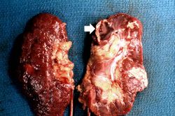 This gross photograph shows a kidney that has been transected longitudinally at autopsy. The cut surface (right) shows several areas of infarction. The most recent infarct is seen at the top left (arrow). The surface of the kidney (left) shows a marked nodularity and roughening from scarring due to chronic hypertension.
 -
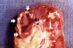 This is a closer view of the triangular-shaped infarction with the base at the cortical surface (1) and the apex at the corticomedullary junction (2).
 -
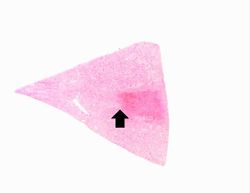 This low-power photomicrograph of the kidney illustrates a sharply demarcated area of red discoloration extending from the capsule to the cortical medullary junction (arrow).
 -
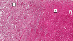 A higher-power photomicrograph shows the edge of this reddish area, illustrating coagulation necrosis (1) compared to the normal tissue (2). The necrotic tubules in this hemorrhagic, red infarct are hypereosinophilic. Compare the tubules on the right with the normal tubules seen in the left-hand portion of the slide. Note the interstitial hemorrhage which is associated with vascular leakage within this necrotic region in the tissue.
 -
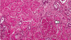 This higher-power view of the infarct demonstrates retention of the tubular structure and cellular outlines. In the lower right-hand corner is a barely identifiable glomerulus (1). Note that, although the cellular architecture is retained, there are no nuclei within the renal tubular cells. The nuclei visible in this photomicrograph are the nuclei of inflammatory cells.
 -
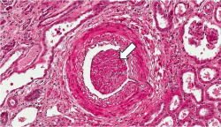 This section, taken at the cortical medullary junction, illustrates a blood vessel in the upper right portion of the slide (arrow) which is filled with thrombotic material. This vessel has occluded an end artery resulting in ischemia and infarction.
 -
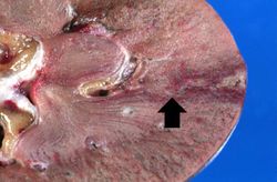 This gross photograph of an infarcted kidney is from another case. The triangular shape of an infarct is prominent on the right side of the image; the apex (arrow) of the triangle is evident at the corticomedullary junction.


Study Questions
Additional Resources
Reference
- eMedicine Medical Library: Renal Cortical Necrosis
- eMedicine Medical Library: Acute Tubular Necrosis
- Merck Manual: Renal Cortical Necrosis
Journal Articles
- Domanovits H, Paulis M, Nikfardjam M, Meron G, Kürkciyan I, Bankier AA, Laggner AN. Acute renal infarction: clinical characteristics of 17 patients. Medicine (Baltimore) 1999 Nov; 78(6): 386-94.
Images
Related IPLab Cases
- Lab 4: Kidney: Atheromatous Emboli
- Lab 5: Kidney: Nodular Intercapillary Glomerulosclerosis
- Lab 6: Kidney: Glomerulonephritis
- Lab 6: Kidney: Acute Transplant Rejection
- Lab 6: Kidney: Chronic Transplant Rejection
| |||||