IPLab:Lab 2:Atrophy: Difference between revisions
Seung Park (talk | contribs) |
|||
| (One intermediate revision by one other user not shown) | |||
| Line 17: | Line 17: | ||
File:IPLab2Atrophy10.jpg|This gross photograph shows a normal brain (left) and a brain from a geriatric patient (right). Note the decreased size, the narrowed gyri, and the widened sulci of the brain from this octogenarian. What is the cause of atrophy in this case? | File:IPLab2Atrophy10.jpg|This gross photograph shows a normal brain (left) and a brain from a geriatric patient (right). Note the decreased size, the narrowed gyri, and the widened sulci of the brain from this octogenarian. What is the cause of atrophy in this case? | ||
</gallery> | </gallery> | ||
== Virtual Microscopy == | |||
=== Testis: Atrophy === | |||
<peir-vm>IPLab2Atrophy</peir-vm> | |||
===Normal Testis === | |||
<peir-vm>UAB-Histology-00045</peir-vm> | |||
== Study Questions == | == Study Questions == | ||
Latest revision as of 14:11, 16 September 2015
Clinical Summary
This 74-year-old man was found to have carcinoma of the prostate six years prior to his death. The serum prostate-specific antigen (PSA) level was 9.7 ng/ml. His plasma acid phosphatase level on repeated tests ranged from 2 to 4 IU/L. X-rays revealed metastatic lesions in the patient's pelvis and vertebral column. He refused to have an orchiectomy and was placed on estrogen therapy (diethylstilbestrol).
The patient's terminal admission was for renal failure. The tumor had grown into the floor of the urinary bladder causing obstruction of the ureters at their entrance to the bladder. The patient developed acute hydronephrosis which was drained by nephrostomies. Eventually, infection developed in the kidneys resulting in Gram-negative sepsis and death.
Images
-
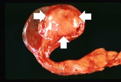 This is a gross photograph of an atrophied testis (arrows).
 -
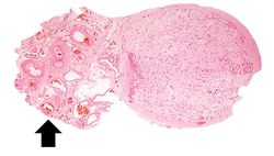 This is a low-power photomicrograph of an atrophic testis. Attached to the testis are several vessels (arrow) which are part of the epididymis and the vas deferens.
 -
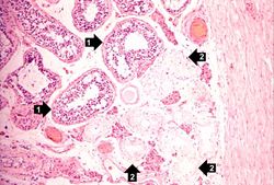 This is a higher-power photomicrograph of an atrophic testis. In this section there are seminiferous tubules with viable cells (1) although there are no visible spermatocytes. Other seminiferous tubules are completely acellular and have a pale pink hyaline appearance (2).
 -
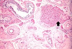 This is a higher-power photomicrograph indicating loss of testicular parenchymal tissue. There are very few recognizable spermatic cells in this tissue. The cluster of cells in the upper right is a focus of interstitial or Leydig cells (arrow). These cells are not affected by the hormone-induced atrophy process.
 -
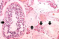 A seminiferous tubule is shown on the left containing remnants of spermatocytes (1). There are no mature sperm present. On the right-hand portion of the slide are remnants of other spermatic tubules which have completely atrophied and lost all of their spermatogenic cells (2). Note the eosinophilic Leydig cells present between the atrophic tubules (3).
 -
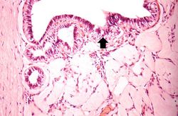 In this slide, the atrophy of the tubules is extending to include the Rete testes (arrow) as well.
 -
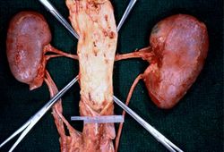 In this gross photograph of kidneys and the abdominal aorta, there is narrowing of the left renal artery at its ostium from the aorta. This atherosclerotic narrowing of the renal artery causes reduced blood pressure in the kidney whose artery is affected. The result is a characteristic Goldblatt’s hypertension syndrome. Note that the kidney with the narrowed renal artery is atrophied compared to the kidney with the normal vessel on the right. What is the cause of the atrophy in this kidney?
 -
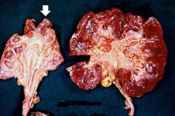 These kidneys were removed from a patient who had blockage of one ureter leading to increased pressure in the renal pelvis. The increased pressure produced hydronephrosis (arrow) in one kidney. What is the cause of atrophy in this case?
 -
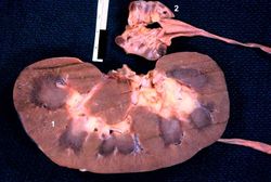 The two kidneys in this slide are from the same patient. One kidney (1) is relatively normal, although increased in size due to compensatory hypertrophy. The other kidney (2) is very small with only rudimentary nodules of renal parenchyma. This kidney had never developed and therefore this process represents hypoplasia. How does one differentiate between atrophy and hypoplasia?
 -
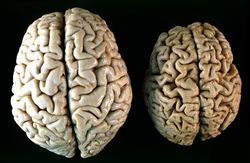 This gross photograph shows a normal brain (left) and a brain from a geriatric patient (right). Note the decreased size, the narrowed gyri, and the widened sulci of the brain from this octogenarian. What is the cause of atrophy in this case?


Virtual Microscopy
Testis: Atrophy
Normal Testis
Study Questions
Additional Resources
Reference
- National Cancer Institute: Prostate Cancer Treatment
- eMedicine Medical Library: Prostate-Specific Antigen
- eMedicine Medical Library: Prostate Cancer
- Merck Manual: Prostate Cancer
Journal Articles
- van Leenders GJ, Schalken JA. Stem cell differentiation within the human prostate epithelium: implications for prostate carcinogenesis. BJU Int 2001 Sep;88 Suppl 2:35-42; discussion 49-50.
Images
| |||||