IPLab:Lab 8:HSV Encephalitis: Difference between revisions
Seung Park (talk | contribs) Created page with "== Images == <gallery heights="250px" widths="250px"> IPLab8HSVEncephalitis1.jpg|This is a gross photograph of a section of brain showing multiple small, punctate hemorrhages ..." |
Seung Park (talk | contribs) No edit summary |
||
| (10 intermediate revisions by 2 users not shown) | |||
| Line 1: | Line 1: | ||
== Clinical Summary == | |||
This 30-year-old white male experienced a generalized tonic-clonic seizure and was subsequently started on a course of Dilantin. He did well, but later developed a headache lasting over a week, which was associated with tonic-clonic seizures, fever, and--toward the end of this period--ataxia. The patient improved and returned to work, but the headache returned. A lumbar puncture was then performed which showed 22 cells/mm³ (all lymphocytes), protein of 88 grams/L, and a glucose level of 49 mg/dL (with a simultaneous serum glucose of 83 mg/dL). These findings were compatible with a viral infection. Despite therapy, the patient had another seizure and again developed fever. At that time, a brain biopsy was performed which showed herpetic encephalitis. Despite aggressive antiviral therapy the patient died. | |||
== Images == | == Images == | ||
<gallery heights="250px" widths="250px"> | <gallery heights="250px" widths="250px"> | ||
| Line 14: | Line 18: | ||
IPLab8HSVEncephalitis12.jpg|This is a photomicrograph of a brain section stained with an antibody against herpes simplex. Even at this magnification, it is easy to pick out cells that are positive for the virus (arrows). | IPLab8HSVEncephalitis12.jpg|This is a photomicrograph of a brain section stained with an antibody against herpes simplex. Even at this magnification, it is easy to pick out cells that are positive for the virus (arrows). | ||
</gallery> | </gallery> | ||
== Virtual Microscopy == | |||
<peir-vm>IPLab8HSVEncephalitis</peir-vm> | |||
== Study Questions == | |||
* <spoiler text="What age groups are susceptible to herpetic encephalitis?">HSV-1 (labialis) produces encephalitis in any age group but is most common in children and young adults. Only about 10% of the patients have a history of prior labial herpes.</spoiler> | |||
* <spoiler text="What type of injury is produced by herpetic encephalitis, what areas of the brain are affected, and what are the most common clinical presenting signs/symptoms?">Encephalitis usually involves the inferior and medial regions of the temporal lobes and the orbital gyri of the frontal lobes. The infection is grossly necrotizing and often hemorrhagic with perivascular inflammatory infiltrates and Cowdry intranuclear viral inclusion bodies in both neurons and glia. The most commonly observed clinical presenting symptoms are alterations in mood, memory, and behavior.</spoiler> | |||
== Additional Resources == | |||
=== Reference === | |||
* [http://emedicine.medscape.com/article/218580-overview eMedicine Medical Library: Herpes Simplex] | |||
* [http://emedicine.medscape.com/article/783113-overview eMedicine Medical Library: Herpes Simplex in Emergency Medicine] | |||
* [http://emedicine.medscape.com/article/1165183-overview eMedicine Medical Library: Herpes Simplex Encephalitis] | |||
* [http://www.merckmanuals.com/professional/infectious_diseases/herpesviruses/herpes_simplex_virus_hsv_infections.html Merck Manual: Herpes Simplex Virus (HSV) Infections] | |||
* [http://www.merckmanuals.com/professional/neurologic_disorders/brain_infections/encephalitis.html Merck Manual: Encephalitis] | |||
=== Journal Articles === | |||
* Edlow JA, Panagos PD, Godwin SA, Thomas TL, Decker WW; American College of Emergency Physicians. [http://www.ncbi.nlm.nih.gov/pubmed/18809105 Clinical policy: critical issues in the evaluation and management of adult patients presenting to the emergency department with acute headache]. ''Ann Emerg Med'' 2008 Oct;52(4):407-36. | |||
* Thomson RB Jr, Bertram H. [http://www.ncbi.nlm.nih.gov/pubmed/11780267 Laboratory diagnosis of central nervous system infections]. ''Infect Dis Clin North Am'' 2001 Dec;15(4):1047-71. | |||
=== Images === | |||
* [{{SERVER}}/library/index.php?/tags/127-herpes PEIR Digital Library: Herpes Images] | |||
* [http://library.med.utah.edu/WebPath/CNSHTML/CNSIDX.html#6 WebPath: CNS Infections] | |||
== Related IPLab Cases == | |||
* [[IPLab:Lab 8:HSV Glossitis|Lab 8: Herpes Glossitis]] | |||
{{IPLab 8}} | {{IPLab 8}} | ||
[[Category: IPLab:Lab 8]] | [[Category: IPLab:Lab 8]] | ||
Latest revision as of 11:28, 3 January 2014
Clinical Summary
This 30-year-old white male experienced a generalized tonic-clonic seizure and was subsequently started on a course of Dilantin. He did well, but later developed a headache lasting over a week, which was associated with tonic-clonic seizures, fever, and--toward the end of this period--ataxia. The patient improved and returned to work, but the headache returned. A lumbar puncture was then performed which showed 22 cells/mm³ (all lymphocytes), protein of 88 grams/L, and a glucose level of 49 mg/dL (with a simultaneous serum glucose of 83 mg/dL). These findings were compatible with a viral infection. Despite therapy, the patient had another seizure and again developed fever. At that time, a brain biopsy was performed which showed herpetic encephalitis. Despite aggressive antiviral therapy the patient died.
Images
-
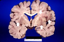 This is a gross photograph of a section of brain showing multiple small, punctate hemorrhages throughout the brain parenchyma (arrows).
 -
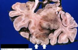 This is a closer view of the previous section of brain showing multiple small, punctate hemorrhages throughout the brain parenchyma (arrows).
 -
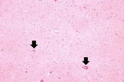 This is a low-power photomicrograph showing a section of brain with numerous perivascular hemorrhages (arrows) and some areas that appear hypercellular.
 -
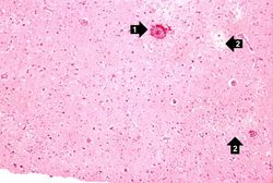 This is a medium-power photomicrograph showing a blood vessel with perivascular hemorrhage (1), areas with loss of brain parenchyma, and edema (2). Even at this power, it can be seen that many of the cells are shrunken and dark red, suggesting that they are necrotic.
 -
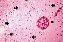 This is a high-power photomicrograph of the previous section. At this power it is easier to see the blood vessel with the perivascular hemorrhage and mild perivascular lymphocytic cuffing (1). In addition, the areas of edema and loss of neurophil (2) can be better appreciated. Red shrunken neurons and glia with pyknotic nuclei (3) are also evident at this power.
 -
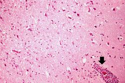 This is another high-power photomicrograph showing a blood vessel with perivascular hemorrhage and mild perivascular lymphocytic cuffing (arrow). In addition, there are numerous red shrunken neurons and glia with pyknotic nuclei throughout this section.
 -
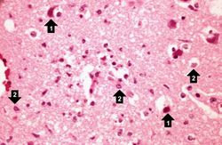 This is a high-power photomicrograph demonstrating clear areas, which indicate edema, and numerous shrunken red necrotic cells (1). At this power, it can be seen that eosinophilic intranuclear inclusion bodies have displaced chromatin to the periphery of the nucleus in some cells (2).
 -
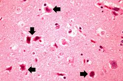 This is a high-power photomicrograph showing several necrotic cells (arrows).
 -
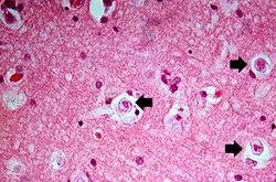 This is a high-power photomicrograph demonstrating cells containing intranuclear inclusion bodies (arrows).
 -
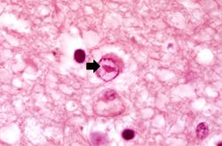 This is another high-power photomicrograph of a cell containing an intranuclear inclusion body (arrow). Note that the nuclear chromatin has been pushed to the outer edges of the nucleus.
 -
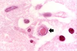 This is another high-power photomicrograph of a cell containing an intranuclear inclusion body (arrow).
 -
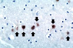 This is a photomicrograph of a brain section stained with an antibody against herpes simplex. Even at this magnification, it is easy to pick out cells that are positive for the virus (arrows).


Virtual Microscopy
Study Questions
Additional Resources
Reference
- eMedicine Medical Library: Herpes Simplex
- eMedicine Medical Library: Herpes Simplex in Emergency Medicine
- eMedicine Medical Library: Herpes Simplex Encephalitis
- Merck Manual: Herpes Simplex Virus (HSV) Infections
- Merck Manual: Encephalitis
Journal Articles
- Edlow JA, Panagos PD, Godwin SA, Thomas TL, Decker WW; American College of Emergency Physicians. Clinical policy: critical issues in the evaluation and management of adult patients presenting to the emergency department with acute headache. Ann Emerg Med 2008 Oct;52(4):407-36.
- Thomson RB Jr, Bertram H. Laboratory diagnosis of central nervous system infections. Infect Dis Clin North Am 2001 Dec;15(4):1047-71.
Images
Related IPLab Cases
| |||||